Wednesday, October 22, 2014

Integumentary System

Down below are some quick notes over the layers of skin and below that is a write-up over an example of what a burn does to your skin. Enjoy.
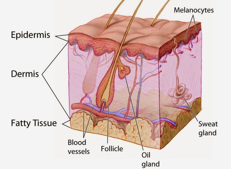
  • Epidermis
    • composed of keratinized stratified squamous epithelium, consisting of 4 distinct cell types and 4 oor 5 layers
    • cell types include keratinocytes, melanocytes, merkel cells, and langerhans cells
    • outer portion of the skin is exposed to the external environment and functions in protection
    • Layer:
      • stratum basal
      • stratum spinosum
      • stratum granulosum
      • stratum lucidum
      • stratum corneum
  • Dermis
    • 2nd major skin region
    • cell types
      • fibroblasts
      • macrophages
      • mast cells
      • white blood cells
    • layers:
      • papillary layer
      • reticular layer
  • Hypodermis
    • subcutaneous layer deep to the skin
    • composed of adipose and areolar connective tissue


The skin consists of three major regions: epidermis, dermis, and hypodermis. Our skin is the first line of defense against pathogens and preventing loss of fluids. When you get burned, the burn is prone to infection.  Severe burns can cause you to become dehydrated due because you burn through all your layers and through all your veins. The layer that functions as waterproofing is the stratum corneum layer.


There are three types of burns. The first burn I'll talk about it first-degree burn. First- degree burn is when the epidermis layer is the only one damaged. When you have a first-degree burn, you'll have redness, swelling, or pain. An example of a first-degree burn is a sunburn or touching a hot or cold object and takes about a week to heal. The next burn is second-degree burn. This burn damages the epidermis and the top layer of the dermis.  The symptoms of second-degree burn mimic first-degree but also blisters appear. The time it takes for these burns to heal depends upon how deep in the dermis the burn is. The third burn is called third-degree burn. This is when the entire thickness of the skin is damaged. Symptoms are the burn appears black, cherry red, or grey-white and there will be no pain due to the nerves being burned. Your skin usually prevents fluid loss but when you have a severe burn, it increases by a great amount. Since your body is losing lots of fluids, you have to replace them at the same rate you're losing them.


There is a way to measure burns, it's called the "rules of nine". This rule estimates the severity of burns. A burn is considered critical if over 25% of the body has second-degree burn, over 20% of the body has third-degree burn, or if there are third-degree burns on the feet, hands, or face. Down below is a link to a website I collected some information from to help me write this post and also I used information off of my teachers powerpoint.


Tuesday, October 21, 2014

Tissue Engineering Techniques


Throughout the years, doctors have came up with new advance ways to do things thanks to all the new technology. Doctors have been focusing on growing body parts for the last twenty years. While they have been studying, they have found out a lot that they want to share with us. Doctors already know how to grow skin for patients that have been burned or have a skin condition where they lose skin.

Their next mission is to be able to grow body parts to replace some time near the future. They hope to be able to grow major body parts and other major functioning assets such as lab-grown cartilage and bone to relieve arthritis suffers and blood vessels, cardiac valves and muscle tissue. Their goal is to make kidneys, corneas, custom-made hearts, livers, breasts, bone marrow and bladders to help patients with diseases or life threatening illnesses. Their first step is to unlock the biochemical signals to influence growth and development. If they add the right combination of compounds, they can coax cells into growing and proliferating. When doing this, scientists need to pay close attention to the physical environment in which cells grow. Scientists can use biodegradable scaffolding to get the organs to look like human organs. By using this technique, cells will have better access to nutrients and waste removal.

Here in the U.S. a lady was in need of a ear and grew one on her arm. When it was done growing, the doctors cut it off and place it where se was missing an ear. Overtime it should look like a natural ear. Hopefully when this tissue engineering is complete, many life threatening illnesses and diseases can be cured and the waiting list for an organ transplant will disappear.


Down below are the sites I gathered my information from.

http://www.pbs.org/saf/1107/features/body.htm

http://gizmodo.com/5946943/woman-grows-a-new-ear-on-her-arm-has-it-attached-to-her-head-warning-graphic

http://www.bbc.co.uk/news/world-asia-china-24282498

Wednesday, October 15, 2014

Histology Lab: Microscopy of Dead Tissue


Down below are pictures that my group took of different kinds of tissues. Located under each picture is a description of the tissue above it. I hope you enjoy. 


Cardiac Muscle:
Description- branching, striated, generally uninucleate cells that interdigitate at specialized junctions 
Function- as it contracts, it propels blood into the circulation 
Location- the walls of the heart 


Skeletal Muscle:
Description- long, cylindrical, multinucleate cells
Function- voluntary movement; locomotion; manipulation of the environment; facial expression; voluntary control
Location- in skeletal muscles attached to bones and occasionally to skin


Puedo-stratified Columnar:
secretion and propulsion of mucus (makes mucus)


Stratified Squamous:
layers of thin epithelium cells, for protection to underlying layers and abration


Nervous Tissue:
Description- neurons are branching cells; cell processes that may be quite long extend from the nucleus-containing cell body; also contributing to nervous tissue are non irritable supporting cells
Function- transmit electrical signals from sensory receptors and the effectors which control their activity
Location- brain, spinal cord, and nerves


Transitional Epithelium:
stretches to permit the dissension of the urinary bladder


Wednesday, October 1, 2014

Human Models of Epithelia


A group of people were put together to demonstrate what the structure of different types of epithelia looks like. Down below are the pictures with the functions, location and definition of each structure.
ENJOY!


Stratified Cuboidal:
Description- Single layer of cube like cells
Location- Kidney tubules; ducts and secretory portions of small glands; ovary surface
Function- Secretion and absorption 


Pseudo-Stratified Cuboidal:
Description- Slightly thicker than squamous
Location- Lines the larger ducts of the mammary glands, sweat glands, and pancreas (protection)


Stratified Columnar:
Description- Single layer of tall cells with round to oval nuclei
Location- Non ciliated type lines most of the digestive tract, gallbladder, and execratory ducts of some glands; ciliated variety lines small bronchi, uterine tubes, and some regions of the uterus 
Function- Absorption; secretion of mucus, enzymes, and other substances; ciliated type propels mucus by ciliary action 


Stratified Squamous:
Description- Thick membrane composed of several cell layers
Location- Nonkeratinized type forms the moist linings of the esophagus, mouth, and vagina 
Function- protects underlying tissues in areas subjected to abrasion


Pseudo-stratified squamous:
Description- Thin cells on top of cells, found on outer layer of the ski 
Location- Oral cavity lining, esophagus, vaginal and anal canal (protection due to many layers)


Pseudo-stratified columnar:
Description- Single layer of cells of differing heights, some not reaching the free surface; nuclei seen at different levels; may contain goblet cells and bear cilia
Location- Non ciliated type in male's sperm-carrying ducts and ducts of large glands; ciliated variety lines the trachea, most of the upper respiratory tract
Function- Secretion, particularly of mucus; propulsion of mucus by ciliary action 


Transitional:
Description- Resembles both stratified squamous and stratified cuboidal; basal cells cuboidal or columnar; surface cells dome shaped or squamous-like, depending on degree of organ stretch
Location- Lines the ureters, bladder, and part of the urethra
Function- Stretches readily and permits dissension of urinary organ by contained urine